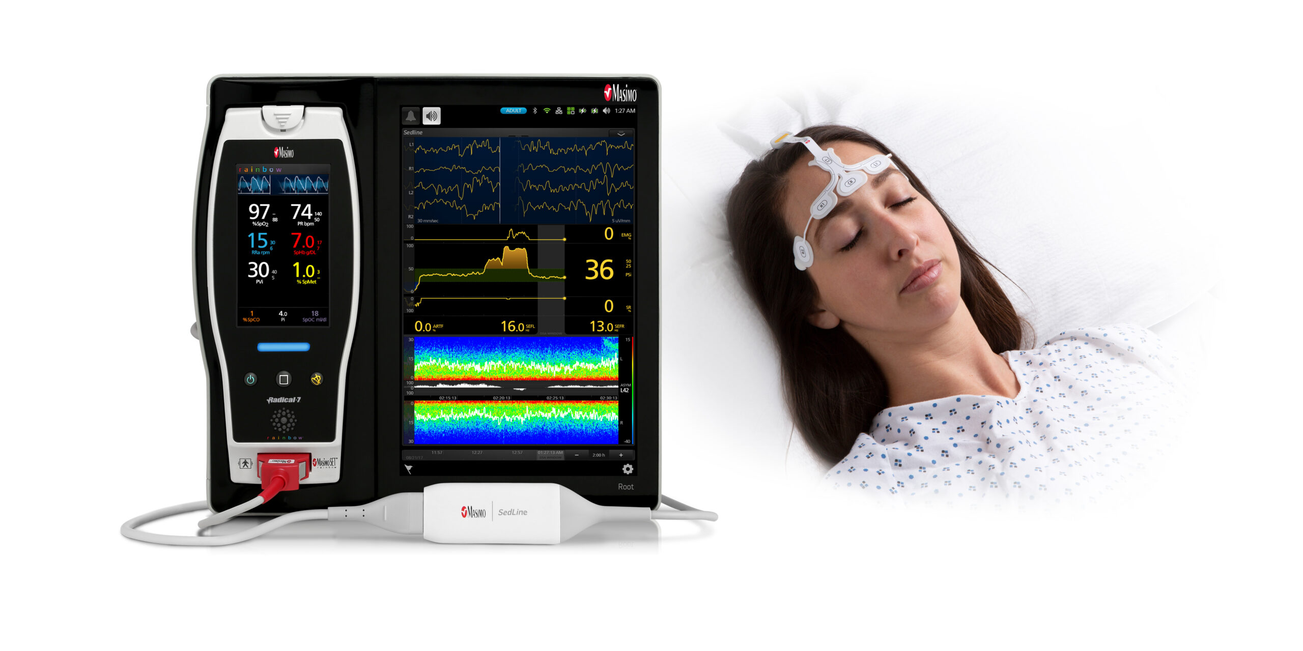
With bilateral electroencephalogram (EEG) signal processing and data acquisition, Root® with Next Generation SedLine Brain Function Monitoring aids clinicians in keeping track of the condition of the brain while it is under anaesthesia.
Features of the upcoming SedLine:
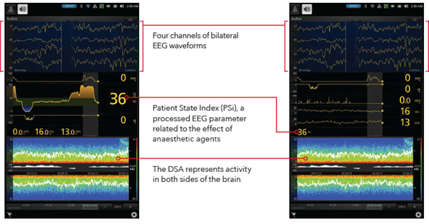
Four frontal EEG waveform channels recorded simultaneously
An improved Patient State Index (PSi), an EEG parameter that has been processed to reflect the impact of anaesthetic medications
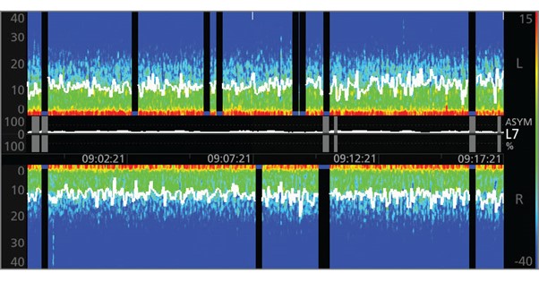
A Density Spectral Array (DSA) display that shows spectrograms for the left and right sides of the brain to show how powerful the EEG is on each side of the brain.
An optional Multitaper DSA that could make EEG characteristics more visible
The increased Patient Sate Index (PSi), a processed EEG parameter associated to the impact of anaesthetic medicines, is provided by the upgraded signal processing engine of Next Generation SedLine.
The Next Generation SedLine PSi outperformed the original SedLine PSi, according to EEG experts. Performance of the Next Generation PSi was determined to have improved by 25% on average.
Next Generation SedLine offers clinicians the flexibility of choosing to display either an enhanced Multitaper Density Spectral Array (DSA) or a standard Hanning DSA. The DSA contains left and right spectrograms representing the power of the EEG on both sides of the brain.
When using a Multitaper DSA, EEG data are transformed into the frequency domain, which may provide a better display of EEG features.
The Masimo Open Connect® (MOC-9®) connections on the Next Generation SedLine module make it simple to connect it to the Root patient monitoring platform. The adaptable, simple-to-understand Root display provides numerous perspectives of brain monitoring data, increasing visibility in the operating room and intensive care unit.
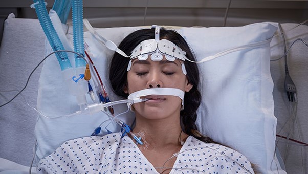
Four active EEG leads collect data in the frontal lobe
Soft foam pads for comfortable application on a patient’s forehead
Adhesive anchors ensure secure sensor placement for optimal signal quality
Pre-filled gel electrodes help streamline sensor application workflows
Application graphics for O3 regional oximetry sensor placement simplify simultaneous application of both monitoring technologies